Pigmented Lesions: Melasma and Moles
Book NowMelasma, Moles and Pigmentation Treatment in Ottawa
What is melasma?
Melasma is a form of hyperpigmentation that is caused and exacerbated by sun damage, genetics, ageing, and trauma. It’s areas of darkened, brown or blue-grey patches or freckle-like spots that become more pronounced with time. It’s an overproduction of cells that is common, harmless and treatable.
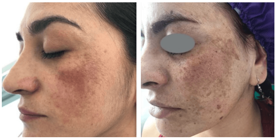
How do you treat melasma and other types of lesions?
At Inovo Medical we own the best technology and are able to treat most types of lesions that you may suffer from. Using the StarWalker Pico Nano laser, we treat your non-raised dermal, epidermal and strongly pigmented lesions using Fotona’s state-of-the art Pico Nano StarWalker laser. This is a pigment selective laser that innovates the treatment of melasma, birthmarks, solar lentigines, and tattoo removal. The laser destroys pigment without burning the skin or stimulating cells to produce more pigments.
It’s the only device that reduces downtime and carries no risks of causing dermal hyperpigmentation, which is usually a concern in Asian, Black, and Middle Eastern skin tones.
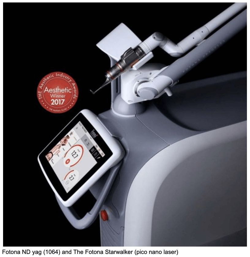
Pico refers to the term picosecond which is one trillionth of a second. The pico laser is the fastest rate picosecond laser on the market. The pulses induce the remodelling of skin without heat making the safest treatment option available.
Often it is recommended to combine your melasma treatment with a skin-bleaching cream (hydroquinone). Treatments such as laser peels (non pico lasers), deep fractionated lasers, strong chemical peels, and IPL are mildly effective and carry the risk of worsening your symptoms.
Moles and Skin Lesions
What are the different types of lesions?
Lesions can be classified into: vascular lesions, dermal lesions, epidermal lesions and subdermal lesions.Each lesion possesses unique characteristics that must be considered when choosing the technology to treat it. Some clinics and spas often suggest treatments on various lesions, like IPL for example, that are ineffective and may even increase symptoms. That’s because they use non specific lasers, trying to do more with less.
CURRENT PROMOTIONS
Buy 1 Laser Mole Session,
Get Your Second One FREE
(OR get 25% off a single session)
Understanding the depth of lesions: Epidermal, Dermal and Subdermal
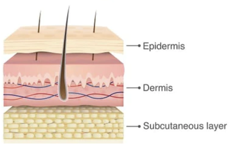
There are three layers of tissue that make up your skin. Dermal, Epidermal and Subdermal (subcutaneous). Epidermal lesions are located on your skin’s top layer. Dermal lesions reach the middle skin and subdermal lesions are deeply situated under the dermis.
Epidermal lesions are mostly caused by sun damage (freckles, solar lentigo, etc.), but also include:
- Warts (hard bumps that can appear on any part of your skin. They start from a virus and are contagious)
- Skin tags (A build up of tissue with a stem where the tag connects to your body and tend to be skin coloured)
- Certain types of moles and nevus in that category
- Seborrheic keratosis (pictured here)
- Milias (multiple small, white bumps (cysts) that are often located on the nose and cheeks
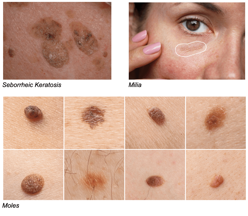
For other epidermal lesions – plane warts, seborrheic keratosis, skin tag, fibrous papule, or milias – we use Fotona’s SP Dynamis Pro laser to remove the lesions one layer at a time.
Dermal lesions refer to lesions that are within the dermis (below epidermis). It’s a post-inflammatory hyperpigmentation caused by acne, scarring, bug bites etc.
Are there lesions that are untreatable?
If there are concerns your lesion(s) may be cancerous we will work together to develop an action plan.
Subdermal lesions are also not treated here. These lesions are deep bumps under your skin that we wouldn’t be able to remove and would require a small surgery.
Such lesions include:
- An abscess
- Sebaceous cysts
- Deep dermal or subdermal nodules
Other dermal pigmented lesions we treat:
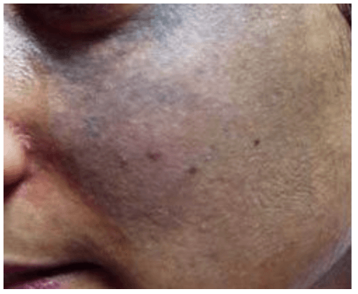
Nevus of Ota
A type of dermal melanocytosis (excessive melanocytes in the tissue) that causes the hyperpigmentation of an eye and the surrounding area. It often takes the form of bluish or brownish pigment around the eye, along with pigment appearing on the whites of the eyes.
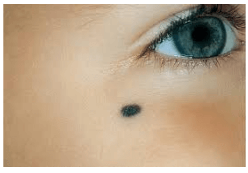
Blue Nevus (mole)
A type of melanocytic nevus. The blue colour is caused by pigment being deeper in the skin than a normal nevus (mole).
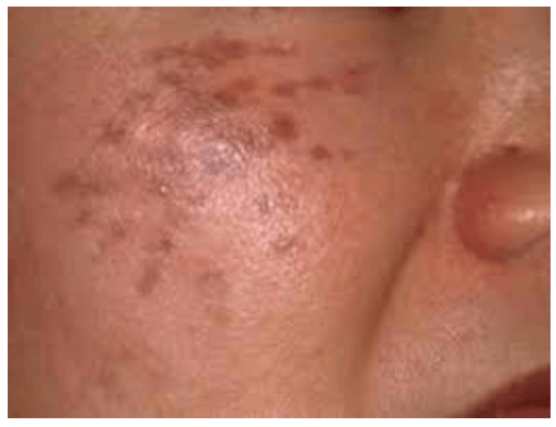
Abnom/Hri’s Nevus
Presents itself as asymptomatic blue-brown or slate grey coloured macules located bilaterally on the face without mucosal involvement. It’s a condition usually found in middle-aged women of Asian descent.
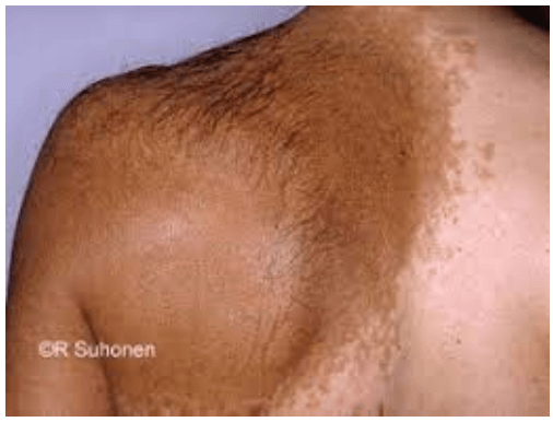
Becker’s Nevus
This is a skin disorder that predominantly affects men. The nevus can be present at birth but more often shows up around puberty. It is also known as pigmented hair nevus.
Other epidermal pigmented lesions we treat:
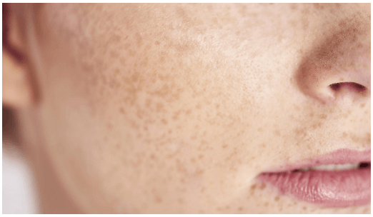
Freckles
A small patch of light brown colour on the skin that is caused by genetics and sun exposure. This exposure often makes freckles become more pronounced.
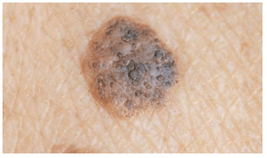
Seborrheic Keratosis
This non-cancerous skin growth tends to come with age. They’re usually black, brown or a light tan. The growth itself looks waxy, scaly and slightly raised. This condition is caused by excessive sun exposure and can be genetically prone to this type of lesion.
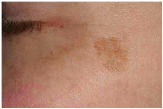
Lentigines/Lentigo
This condition is common over the age of 40 and is marked by small brown patches on the skin that are light brown and caused by exposure to the sun. The more exposure, the darker they get.
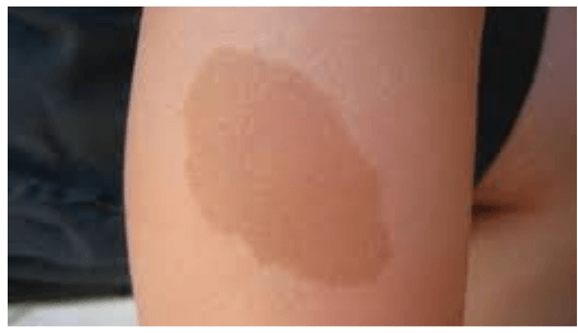
Cafe au Lait
A type of birthmark characterized by flat patches on the skin that vary in size. They’re light brown and darken with sun exposure. They are distinct due to their irregular edges and colour variation. If removed they can return within a year.
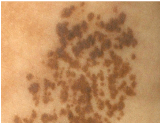
Nevus Spilus
A lesion (speckled lentiginous lesion nevus) that presents as light brown or tan macules speckled with smaller, darker macules or papules. Caused by genetics or acquired. There is potential for this to turn into melanoma and a biopsy is recommended before any sort or treatment or removal.
How are different types of lesions treated?
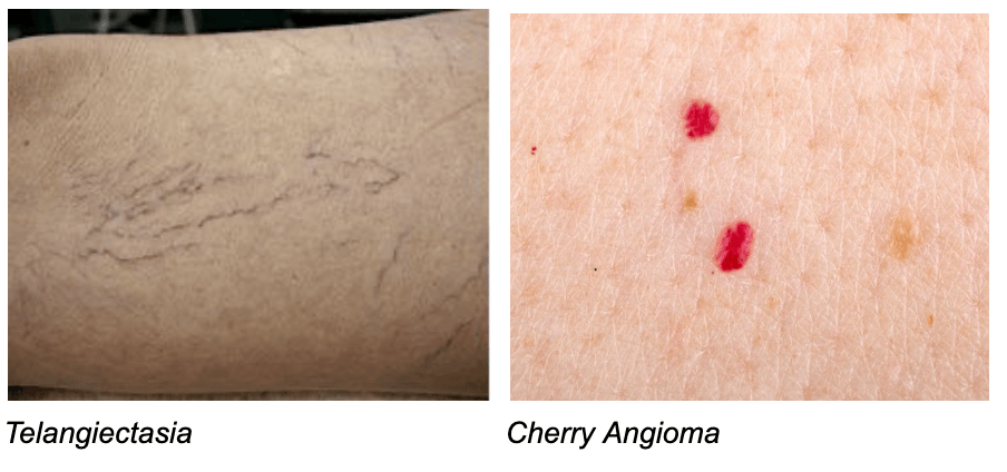
Vascular lesions like rosacea telangiectasias, hemangioma, spider angioma, cherry angiomas and other vascular lesions, are best treated with the Fotona SP Dynamis and the Fotona StarWalker laser. For more information on vascular lesion treatments, click here.
During Your Consultation
- Our cosmetic medical staff will meet with you.
- We’ll go over your unique situation and your goals.
- We’ll discuss what to realistically look forward to.
- We’ll present the various treatment options available.
- We’ll recommend what we feel meet your needs best.
- Finally, if you’re ready we’ll book your treatment.


